Reports tied to this Journal
Author
|
Message

|
 Culture Journal, Species: Thalassiosira weissflogii
Wednesday, August 31, 2011 1:23 AM
( permalink)
Culturing Journal DataSheet
This first post should be updated regularly to include new information as events take place or changes are made to your system General Species: Thalassiosira weissflogii Species description: A large diatom (6-20µm x 8-15µm) Culture source (link if possible ): Another MBI user, WDT. If algae, CCMP # (Optional ): CCMP1051 http://ccmp.bigelow.edu/ Culture Establishment Date: 8/17/2011 Continuation Date: Culturing Vessel Details Salinity: 1.020 Temperature: Low 70s F pH: Not measured
Vessel description: Initially, a 1000 ml Erlenmeyer flask, due to the small size of the starter. Subsequently, 2000 ml borosilicate media jars. Lighting description: 2 x T5 21 watt 6500K fluorescent bulbs. Lighting cycle: 18 H on, 6 H off Aeration description: Moderate aeration with a rigid airline and a filtered air source. Methodologies Split methodology: Culture medium description: Guillard's F/2 medium in the form of FAF Micro Algae Grow, supplemented with sodium silicate. Cell count: Not measured yet. Reference links: Additional Information (No Pictures or Videos in the Section Please) Notes: You will be required to provide photographic evidence and as much detail as possible about your project in this thread.
If your thread does not contain detailed enough photos and information the MBI Council will not be able to approve your reports.
|
|
|
 Re:Culture Journal, Species: Thalassiosira weissflogii
Thursday, September 1, 2011 10:26 PM
( permalink)
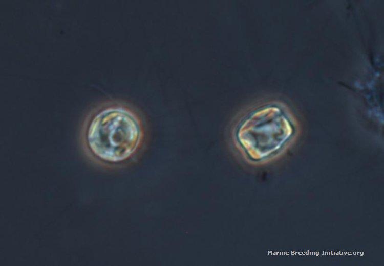
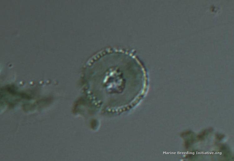
Today was the first split of the first 2000 ml bottle of the Thalassiosira. For images documenting this, I took a sample into the lab to get some microscope images of the culture, to confirm it is the diatom Thalassiosira, and not just some contaminate. Thalassiosira is a disc-shaped diatom, approximately 6-20 microns across. The photos below show two different magnifications and two different treatments. The first photo is taken at 400x. A drop of dense culture was placed on a clean slide, and a cover slip was placed on it. There are two diatoms in this image, one viewed dorsally, and one viewed laterally. If you look carefully, you can see the fine setae radiating out from both diatoms.  The second photo is taken at 1000x. Appx. 1 ml of dense culture was placed into a 1 ml vial, and was centrifuged. The supernatant was poured off. The vial was refilled with ethanol, mixed, and centrifuged again. The supernatant was poured off, and replaced with fresh ethanol. A drop from the resulting contents of the vial was placed onto a clean slide, and allowed to evaporate. It was then examined at 1000x using an oil immersion lens. While this means of observing reveals much more fine detail, the rough treatment during centrifugation apparently removed all the fine setae, and seems to have othewise damaged the cells. The microbiologists at work admit that they are unaccustomed to dealing with fragile, larger organisms like diatoms; they usually are handling bacteria and yeasts. 
|
|
|
 Re:Culture Journal, Species: Thalassiosira weissflogii
Friday, September 2, 2011 9:35 PM
( permalink)
amazing pictures! What microscope did you use?
|
|
|
 Re:Culture Journal, Species: Thalassiosira weissflogii
Friday, September 2, 2011 10:38 PM
( permalink)
Thanks! It's an Olympus BX60.
|
|
|
 Re:Culture Journal, Species: Thalassiosira weissflogii
Saturday, September 3, 2011 8:26 AM
( permalink)
Great shots!!
Not sure if it matters, but my source was carolina.com
|
|
|
 Re:Culture Journal, Species: Thalassiosira weissflogii
Saturday, September 3, 2011 10:39 AM
( permalink)
Thanks again, wdt, for the diatom starter, the copepods, and the rotifers. All three are doing very well for me!
|
|
|
|